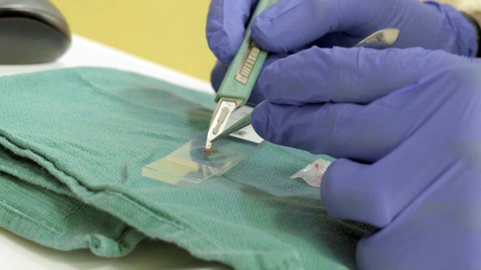
Brain surgeons are bringing artificial intelligence and new imaging techniques into the operating room, to diagnose tumors as accurately as pathologists, and much faster, according to a report in the journal Nature Medicine.
The new approach streamlines the standard practice of analyzing tissue samples while the patient is still on the operating table, to help guide brain surgery and later treatment.
The traditional method, which requires sending the tissue to a lab, freezing and staining it, then peering at it through a microscope, takes 20 to 30 minutes or longer. The new technique takes two and a half minutes. Like the old method, it requires that tissue be removed from the brain, but uses lasers to create images and a computer to read them in the operating room.
“Although we often have clues based on preoperative M.R.I., establishing diagnosis is a primary goal of almost all brain tumor operations, whether we’re removing a tumor or just taking a biopsy,” said Dr. Daniel A. Orringer, a neurosurgeon at N.Y.U. Langone Health and the senior author of the report.
In addition to speeding up the process, the new technique can also detect some details that traditional methods may miss, like the spread of a tumor along nerve fibers, he said. And unlike the usual method, the new one does not destroy the sample, so the tissue can be used again for further testing.
The new process may also help in other procedures where doctors need to analyze tissue while they are still operating, such as head and neck, breast, skin and gynecologic surgery, the report said. It also noted that there is a shortage of neuropathologists, and suggested that the new technology might help fill the gap in medical centers that lack the specialty.

Algorithms are also being developed to help detect lung cancers on CT scans, diagnose eye disease in people with diabetes and find cancer on microscope slides. The new report brings artificial intelligence — so-called deep neural networks — a step closer to patients and their treatment.
The study involved brain tissue from 278 patients, analyzed while the surgery was still going on. Each sample was split, with half going to A.I. and half to a neuropathologist. The diagnoses were later judged right or wrong based on whether they agreed with the findings of lengthier and more extensive tests performed after the surgery.
The result was a draw: humans, 93.9 percent correct; A.I., 94.6 percent.
The study was paid for by the National Cancer Institute, the University of Michigan and private foundations. Dr. Orringer owns stock in the company that made the imaging system, as do several co-authors, who are company employees. He conducted the research at the University of Michigan, before moving to New York.
“Having an accurate intra-operative diagnosis is going to be very useful,” said Dr. Joshua Bederson, the chairman of neurosurgery for the Mount Sinai Health System, who was not involved in the study. He added, “I think they understated the significance of this.”
He said the traditional method of examining tissue during brain surgery, called a frozen section, often took much longer than 30 minutes, and was often far less accurate than it was in the study. At some centers, he said, brain surgeons do not even order frozen sections because they do not trust them and prefer to wait for tissue processing after the surgery, which may take weeks to complete.
“The neuropathologists I work with are outstanding,” Dr. Bederson said. “They hate frozen sections. They don’t want us to make life-altering decisions based on something that’s not so reliable.”
Dr. Bederson said that the study authors had set a very high bar for their new technique by pitting it against experts at three medical centers renowned for excellence in neurosurgery and neuropathology: Columbia University in New York, the University of Miami and the University of Michigan, Ann Arbor.
“I think that what happened with this study is that because they wanted to do a good comparison, they had the best of the best of the traditional method, which I think far exceeds what’s available in most cases,” Dr. Bederson said.
The key to the study was the use of lasers to scan tissue samples with certain wavelengths of light, a technique called stimulated Raman histology. Different types of tissue scatter the light in distinctive ways. The light hits a detector, which emits a signal that a computer can process to reconstruct the image and identify the tissue.
The system also generates virtual images similar to traditional slides that humans can examine.
The researchers used images from tissue from 415 brain surgery patients to train an artificial intelligence system to identify the 10 most common types of brain tumor.
Some types of brain tumor are so rare that there is not enough data on them to train an A.I. system, so the system in the study was designed to essentially toss out samples it could not identify.
Over all, the system did make mistakes: It misdiagnosed 14 cases that the humans got right. And the doctors missed 17 cases that the computer got right.
“I couldn’t have hoped for a better result,” Dr. Orringer said. “It’s exciting. It says the combination of an algorithm plus human intuition improves our ability to predict diagnosis.”
In his own practice, Dr. Orringer said that he often used the system to determine quickly whether he had removed as much of a brain tumor as possible, or should keep cutting.
“If I have six questions during an operation, I can get them answered without having six times 30 or 40 minutes,” he said. “I didn’t do this before. It’s a lot of burden to the patient to be under anesthesia for that long.”
Dr. Bederson said that he had participated in a pilot study of a system similar to the one in the study and wanted to use it, and that his hospital was considering acquiring the technology.
“It won’t change brain surgery,” he said, “but it’s going to add a significant new tool, more significant than they’ve stated.”
[Like the Science Times page on Facebook. | Sign up for the Science Times newsletter.]
