
By SAURABH JHA and JEANNE ELKIN
Mr. Smith’s pneumonia was clinically shy. He didn’t have a fever. His white blood cells hadn’t increased. The only sign of an infection, other than his cough, was that his lung wasn’t as dark as it should be on the radiograph. The radiologist, taught to see, noticed that the normally crisp border between the heart and the lung was blurred like ink smudged on blotting paper. Something that had colonized the lungs was stopping the x-rays.
Hundred and twenty-five years ago, Wilhelm Conrad Roentgen, a German physicist and the Rector at the University of Wurzburg, made an accidental discovery by seeing something he wasn’t watching. Roentgen was studying cathode rays – invisible forces created by electricity. Using a Crookes tube, a pear-shaped vacuum glass tube with a pair of electrodes, Roentgen would fire the cathode rays from one end by an electric jolt. At the other end, the rays would leave the tube through a small hole, and generate colorful light on striking fluorescent material placed near the tube.
By then photography and fluorescence had captured literary and scientific imagination. In Arthur Conan Doyle’s Hound of the Baskervilles, the fire-breathing dog’s jaw had been drenched in phosphorus by its owner. Electricity and magnetism were the new forces. Physicists were experimenting in the backwaters of the electromagnetic spectrum without knowing where they were.
On November 8th, 1895, when after supper Roentgen went to his laboratory for routine experiments, something else caught Roentgen’s eyes. Roentgen closed the curtains. He wanted his pupils maximally dilated to spot tiny flickers of light. When he turned the voltage on the Crookes tube, he noticed that a paper soaked in barium platinocyanide on a bench nine feet away flickered. Cathode rays traveled only a few centimeters. Also, he had covered the tube with heavy cardboard to stop light. Why then did the paper glow?
Roentgen’s scientific ethos was replication. Only reproducibility of results could convince him that his eyes weren’t being deceived. The glow wasn’t an artifact because the paper glowed only when the Crooke’s tube was on. Had he underestimated the distance cathode rays travelled? The paper still glowed when placed further away from the tube. Not even a deck of cards stopped the glow.
By the nineteenth century, physicists had replaced the chemists as nature’s alchemists. Whereas the chemists tried converting metal to gold, physicists created invisible material out of invisible material. One such invisible material were cathode rays, which later turned out to be electrons. Roentgen didn’t know their precise nature. He inferred their properties only by careful experimentation. He didn’t think they were responsible for the distant glow. But cathode rays were somehow responsible for this “new kind of rays”, which were similar to light but could penetrate solid material, which he called “x-rays” – the “x” denoting that their nature was unknown.
X-rays were discovered accidentally because Roentgen left the fluorescent paper on the distant bench accidentally. But they had been discovered because Roentgen followed his sharp observation with diligent investigation. X-rays might have been discovered sooner had they been noticed. A few years earlier, Arthur Godspeed, a physicist at the University of Pennsylvania, who also experimented with cathode rays, developed photos, some of which had disc-shaped shadows which, unbeknownst, were cast by coins in the path of x-rays. William Crookes, the inventor of the Crookes tube, returned several photographic plates to the manufacturers because they were fogged. After Roentgen’s discovery, he realized that the “damage” he had reported was actually x-rays’ signature.
Roentgen’s laboratory, like Victor Frankenstein’s study, was a Gothic room with contraptions powered by flashes of electricity. Like Frankenstein, he was unleashing science to unravel the supernatural. With curiosity and doubt, Roentgen spent several weeks experimenting with x-rays. He studied the stopping properties of various metals by observing their shadows. Then on one occasion, when he held a piece of lead in his hand, he saw shadows of his bones.
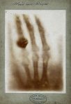
Radiology was conceived on December 22nd, 1895 when Roentgen placed his wife’s left hand in the path of the x-rays. After a fifteen-minute exposure, an iconic photograph emerged. Only bones and a wedding ring cast a shadow. Hitherto, bones were seen only when the dead were opened. Seeing the bones stripped of flesh, naked bones, in the living must have seemed supernatural. Six days later, Roentgen published his findings in a paper titled “On a New Kind of Rays” in the Proceedings of the Wurzburg Physical Medical Society, a relatively obscure publication with rapid publishing time. Roentgen said to Bertha Ludwig, his wife, “now the devil will be to pay.”
On January 5th, 1896 the Vienna Presse, an Austrian Newspaper, published Roentgen’s discovery. Even though there was no social media, and only the telegraph shortened distances, news of his discovery became viral. Within ten days, the London Standard and the New York Times ran the story. Science spread by media. The medical journals came late to the party. The press instantly recognized the medical importance of x-rays, noting that they could photograph broken bones and bullets in human bodies.
The early x-ray enthusiasts were photographers, physicians, engineers such as Thomas Edison, and con artists. X-rays quickly became known for their nefarious potential and became antithetical to Victorian sensibilities. An editorial in the London-based Pall Mall Gazette in March 1896 expressed concern with the “Roentgen Rays” and, cautioning readers of the “revolting indecency” of being able to see people’s bones with the naked eyes, asked that legislature restrict x-ray vision.
The first medical x-ray in the US was of a Colles’ fracture in a boy who injured his wrist, taken by Edwin Frost, a Dartmouth astronomer in February 1896, for his physician brother. In the same month John Cox from McGill University, Canada, localized a bullet in the leg of a patient that had eluded the surgeons. By the end of the year, x-rays joined the battlefield in the Nile expedition, to help surgeons deal with wartime injuries. Much of radiographic practice today can be traced to 1896, the year clinical radiology was born.
Before Roentgen, disease was inferred by sound, by percussion and auscultation. X-rays ensured that disease wasn’t just heard but seen. The Frankfurter Zeitung called x-rays “an epoch-making result of research in exact science.” Befittingly, the first Nobel Prize for Physics, awarded to Roentgen in 1901, was for a medical breakthrough. Roentgen made medicine an applied science by bringing physics into medicine. Later, x-rays unraveled another mystery – the structure of DNA – through x-ray crystallography.
Roentgen dodged fame and declined fortune. He refused to copyright his discovery. He donated his Nobel Prize money to academia. Unassuming in fame, he was unflappable even before his fame. Known for his scout’s honor, he once was expelled from school for refusing to snitch his classmate who had drawn a cartoon of a teacher.
When asked by a journalist what he thought when he saw the glow from the distant paper, he replied, curtly. “I did not think, I investigated.” Roentgen’s legacy to humanity was making disease more visible to doctors. His bequeathal to doctors was the twin act of observation and investigation.
Further Reading
Farmelo G. The Discovery of X-Rays. Scientific American 1995; 11: 86-91
Brailsford JS. Roentgen’s Discovery of X-Rays – Their Application to Medicine and Surgery.” BJR 1946; 19: 453-461
Roentgen WC. On a New Kind of Rays. Translation from German to English of Roentgen’s first X-ray publication by A. Stanton. Science 1896; 3: 227 – 231
Saurabh Jha is a radiologist and contributor to THCB. Jeanne Elkin is MS-3 at the Perelman School of Medicine of the University of Pennsylvania
An edited version of the piece was published in Medscape
Spread the love